Advancements in 3D Printing for Heart Components
Significance of Cardiovascular Research
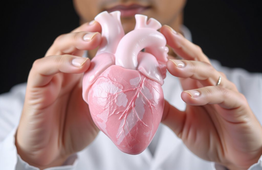
A team of scientists has successfully utilized 3D printing technology to create components of a human heart. Cardiovascular disease continues to be a major cause of morbidity and mortality globally. Research indicates that the prevalence of heart disease is expected to rise as the global population ages. Therefore, ongoing research is essential to discover new strategies for preventing and treating cardiac diseases, particularly for patients experiencing end-stage organ failure who are at high risk of mortality without timely transplants.
The Role of 3D Bioprinting in Medicine
3D bioprinting has revolutionized the production of biomaterials, including implantable meshes that offer therapeutic benefits and serve as research tools. Researchers aspire to utilize bioprinting technology to print cells, biomaterials, and potentially entire organs outside the body. These biomaterials can be subsequently implanted into patients as treatments for various ailments. Recent advancements in 3D bioprinting have enabled the successful creation of patterned tissues, blood vessel-like networks, and other implantable scaffolds.
Challenges in Printing Living Cells
Despite these advancements, the technology has faced limitations in printing living cells and soft biomaterials such as extracellular matrix proteins that are crucial to the heart’s muscle structure. The extracellular matrix serves as a scaffold for cardiomyocytes, the heart’s contractile cells. Printing these proteins, particularly collagen, has presented challenges, as these soft and dynamic biological materials often sag and become unsuitable for further use during the printing process.
Innovative Solutions from Carnegie Mellon University
A research team from Carnegie Mellon University has recently developed an enhanced version of the freeform reversible embedding of suspended hydrogels (FRESH) system. This system successfully prints collagen scaffolds and other soft biomaterials to create various components of the human heart, including blood vessels, heart valves, and cardiac muscle tissue. The new technology employs a temporary support gel to prevent sagging of soft biomaterials. Additionally, the researchers introduced a method to rapidly alter the acidity of the surrounding environment, promoting collagen protein self-assembly to maintain its functional properties.
Successful Creation of Heart Muscle
Beyond producing smaller heart components, the researchers have also generated functional heart muscle using human cardiomyocytes. Evaluations of the 3D printed muscle revealed that the cells exhibited full functionality and displayed the electrical characteristics typical of a healthy heart. Although the goal of 3D printing complete, anatomically accurate, and functional organs is still a challenging endeavor, this study marks a significant advancement. The successful printing of collagen scaffolds and other biomaterials holds promise for developing innovative implantable meshes to address various organ complications.
Conclusion
The research conducted at Carnegie Mellon University represents a critical step forward in the field of tissue engineering and organ regeneration. The implications of these findings extend beyond cardiac applications, potentially impacting numerous areas of medical science.
Reference
Lee, A., Hudson, A. R., Shiwarski, D. J., Tashman, J. W., Hinton, T. J., Yerneni, S., … & Feinberg, A. W. (2019). 3D bioprinting of collagen to rebuild components of the human heart. Science, 365(6452), 482-487.
Image Credit
Image by krzysztof-m from Pixabay