Anatomy and Workings of the Heart
Understanding Heart Anatomy
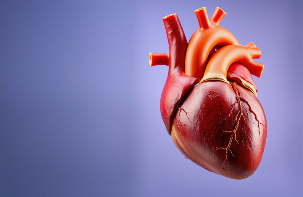
To comprehend the disease processes affecting the heart, it is essential to examine its fundamental anatomy and functionality. The heart is roughly the size of a fist and is positioned beneath the sternum (breastbone), resting on the diaphragm, the primary muscle responsible for breathing. As the most vital muscle in the human body, the heart consists of two sides: the right and left, and contains four chambers. The upper chambers are known as the atria, while the lower chambers are referred to as the ventricles. A muscular wall called the septum divides the two sides. Additionally, the heart features important valves that regulate the one-way flow of blood through the chambers and blood vessels.
Right and Left Side of the Heart
The right side of the heart is responsible for receiving blood from the body and sending it to the lungs for oxygenation. In contrast, the left side of the heart receives oxygen-rich blood from the lungs and pumps it throughout the entire body. The myocardium constitutes the majority of the heart’s mass, playing a crucial role in the pumping of blood throughout both the heart and body.
Circulatory System Overview
Blood is circulated throughout the body via blood vessels, which function as a delivery system. There are three main types of blood vessels:
– **Arteries**: Carry oxygen-rich blood away from the heart.
– **Capillaries**: Serve as exchange vessels that connect arteries to veins.
– **Veins**: Transport deoxygenated blood back toward the heart.
The Heart’s Electrical System
The heart has its own electrical system that synchronizes the work of its chambers, regulating heart rhythm and controlling the frequency of beats. Any issues within this electrical system can lead to arrhythmias, characterized by uncoordinated or random beats, or conditions where the heart beats too quickly (tachycardia) or too slowly (bradycardia).
Coronary Arteries
To function effectively, the heart requires a robust blood supply, which is provided by the coronary arteries. These arteries branch off from the aorta—the large artery that distributes blood to the body from the left ventricle—ensuring that the heart muscle receives the necessary oxygen and nutrients.
Additional Resources
For further learning, consider watching a video on how to take your pulse, or explore more about heart health through Heart 2 Heart initiatives.