The Zika Virus Epidemic and Its Impact
Overview of the Outbreak
The Zika virus gained significant attention during the outbreaks of 2015 and 2016, which affected Brazil, neighboring countries, various Caribbean islands, parts of North America, and several Pacific islands. By February 2016, just one month after the World Health Organization declared Zika an epidemic, it became evident that the virus not only caused symptoms similar to dengue fever—such as fever, rash, and joint pain—but was also linked to severe birth defects.
Research on Ultrasound Use
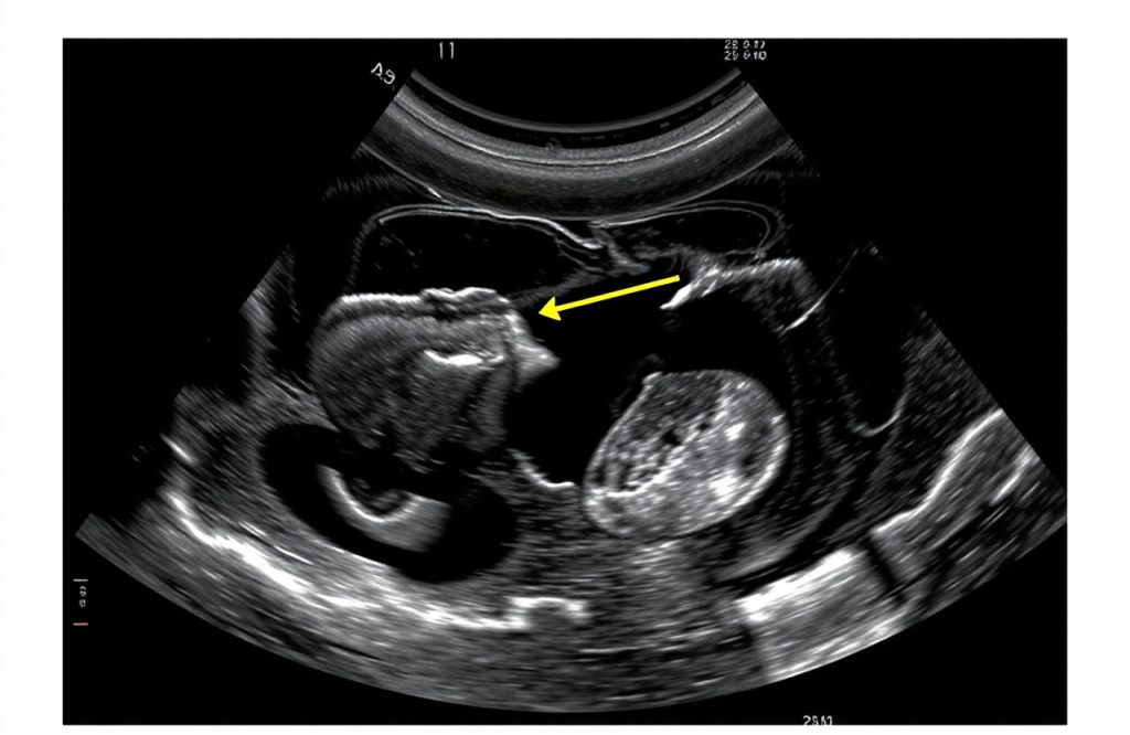
An article published in The Lancet: Child & Adolescent Health highlights research conducted in Martinique focused on the role of ultrasound in identifying brain damage associated with Zika Virus Syndrome. Ultrasound employs sound waves to produce images and is a safe method for examining various fetal conditions, including the brain and hips.
Transmission and Congenital Defects
The Zika virus is transmitted primarily by several species of Aedes mosquitoes, which are also known carriers of dengue fever, yellow fever, and chikungunya. It leads to multiple congenital defects, the most recognized being microcephaly, characterized by an abnormally small head size in infants. However, this study aimed to uncover additional structural defects using ultrasound technology.
Study Details and Findings
The study evaluated fourteen pregnant women who had confirmed Zika virus infections during their first or early second trimester and exhibited abnormal fetal findings. A total of 31 ultrasound images were analyzed for brain abnormalities at various stages of pregnancy. The findings indicated a trend of increasing numbers and severity of anomalies as the pregnancy progressed. Notable issues included:
– Enlarged cerebral ventricles in 10 fetuses, known as ventriculomegaly or hydrocephalus, which can hinder normal brain development.
– General wasting of the brain’s cortex identified in 11 fetuses.
– Calcium deposits observed in 10 fetuses.
– Defects in the corpus callosum, the bundle of neurons connecting the brain’s two hemispheres, found in 10 fetuses.
Interestingly, only 9 fetuses presented with microcephaly, suggesting that other structural abnormalities may be more effective indicators of Zika-related birth defects.
Implications for Screening and Future Research
The researchers concluded that ultrasound is a valuable tool for screening pregnant women who may have been exposed to the Zika virus. They recommended that ultrasounds be conducted between 22 and 26 weeks of pregnancy, emphasizing the need to focus on the identified structural defects. This study represents a critical advancement in developing an efficient strategy for early identification of fetal anomalies linked to Zika virus exposure. Moving forward, research should emphasize strategies for preventing the rapid spread of the Zika virus, given its potential risks to public health.
Written By: Clifton Lewis
References
Ultrasound imaging for identification of cerebral damage in congenital Zika virus syndrome: a case series
Schaub, Bruno et al. The Lancet: Child & Adolescent Health.
http://dx.doi.org/10.1016/S2352-4642(17)30001-9
Add to Flipboard Magazine.