Understanding Phosphorylation in Proteins
The Role of Phosphorylation
Phosphorylation, the addition of a phosphate group to a protein, is crucial for cellular control over various activities. These include signal transmission, energy metabolism, cellular growth, and programmed cell death (apoptosis). This modification occurs post-synthesis, facilitated by enzymes known as protein kinases, which attach phosphate groups to specific amino acids such as serine, threonine, or tyrosine. Conversely, phosphatases are responsible for removing these phosphate groups.
Detecting Phosphorylated Proteins with Western Blotting
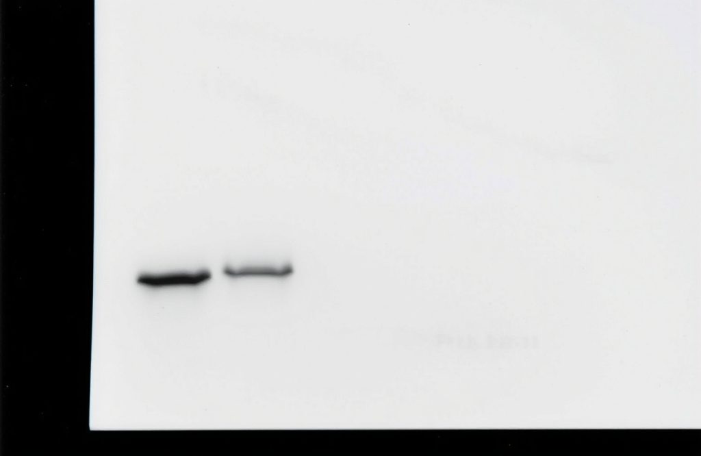
To determine if a protein is phosphorylated, scientists frequently employ a technique called Western blotting. Although the fundamental steps remain consistent, detecting phosphorylated proteins requires careful consideration. A critical component is the use of phospho-specific antibodies, which should be designed to target only the phosphorylated variant of the protein, ensuring accurate experimental results.
Key Considerations for Detecting Phosphorylation
1. Timing of Phosphorylation
Phosphorylation does not occur continuously; it is often triggered by specific conditions, such as exposure to particular signals, disease states, or chemical treatments. To identify when your protein of interest is phosphorylated, consult relevant scientific literature. This research will guide you in selecting appropriate experimental conditions. Conducting time-course experiments can also clarify the duration of phosphorylation, helping you determine the optimal sampling time.
2. Protecting Phosphorylated Proteins
During cell lysis, natural phosphatases can be released, which may quickly remove phosphate groups from proteins, jeopardizing your experiment’s integrity. To mitigate this risk, it is essential to add phosphatase inhibitors to your lysis buffer and include protease inhibitors to prevent degradation. After collection, promptly mix proteins with SDS sample buffer to halt enzyme activity and preserve protein integrity for storage.
3. Selecting the Appropriate Blocking Solution
Blocking is a vital step in Western blotting that prevents antibodies from binding nonspecifically to the membrane. While 5% non-fat milk is commonly used for its availability and cost-effectiveness, it contains casein—a phosphorylated protein that may be detected by anti-phospho antibodies, resulting in elevated background signals. If using milk leads to high background noise, consider switching to BSA (bovine serum albumin), which lacks phosphoproteins.
4. Avoiding Phosphate-Based Buffers
Certain buffers, such as PBS (phosphate-buffered saline), contain phosphate that can interfere with the detection capabilities of anti-phospho antibodies. To avoid this issue, utilize Tris-based buffers such as TBST (Tris-buffered saline with Tween-20) during Western blot procedures. If PBS must be employed at any stage, ensure thorough washing of the membrane with TBST prior to antibody application.
5. Using Specific Antibodies
It is crucial to select antibodies that specifically recognize the phosphorylated form of your protein, avoiding those that may detect both phosphorylated and non-phosphorylated variants. This specificity is particularly vital when analyzing complex samples, such as those from infectious disease models, where accurate detection of modified proteins is essential for understanding disease processes.
6. Assessing Total Protein Levels
Variations in detected phosphorylated protein levels can stem from several factors, including treatment variations or inconsistencies in protein loading. To control for these variables, measure the total amount of the protein (both phosphorylated and non-phosphorylated). This metric serves as a control, allowing you to calculate the percentage of the protein that is phosphorylated. Using PVDF membranes is advisable over nitrocellulose, as they are more robust and suitable for stripping and reusing.
7. Incorporating Proper Controls
Including control samples in your experiments is essential. These should consist of a positive control, where phosphorylation is expected, and a negative control, where phosphorylation should not occur. Additionally, treating samples with phosphatases can confirm the authenticity of your results; if the phosphorylated band disappears post-treatment, it validates that the original band represented a true phosphorylated protein.
8. Enhancing Detection of Low Abundance Phosphoproteins
Detecting phosphorylated proteins can be challenging due to their typically low abundance. To enhance signal detection, consider loading more protein onto the gel by reducing the volume of lysis buffer used. Employ high-sensitivity detection methods, such as enhanced chemiluminescence, and enrich your sample by isolating phosphoproteins through immunoprecipitation before proceeding with Western blotting. Ensure that cells are adequately treated to induce phosphorylation, as noted in previous tips.
9. Utilizing Fluorescent Detection
Stripping and reusing membranes to measure both total and phosphorylated proteins can compromise sample integrity. To avoid this issue, fluorescent Western blotting allows for simultaneous detection of both forms without membrane stripping. This technique involves using two different antibodies—each from separate species—one targeting the phosphorylated form and another for the total protein. Employing fluorescently labeled secondary antibodies for detection yields cleaner results and facilitates more accurate comparisons.
Conclusion
Detecting and analyzing phosphorylated proteins is a complex but crucial aspect of cellular biology. By following these guidelines, researchers can enhance their experimental outcomes and contribute valuable insights into protein function and regulation.