Understanding Cardiovascular Disease Origins
Fetal Development and Cardiovascular Disease
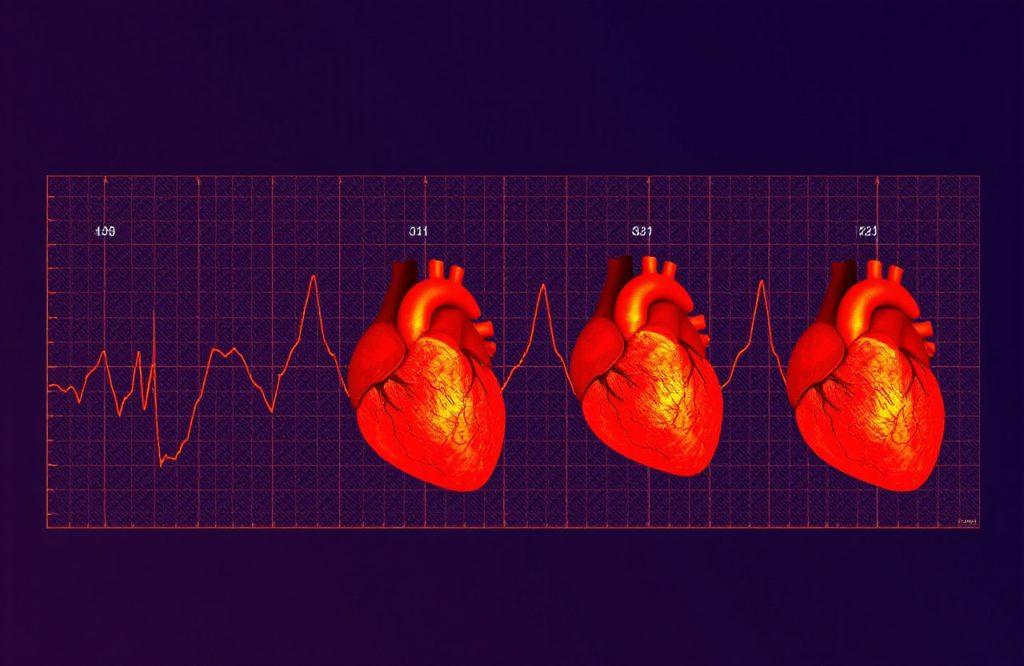
When considering potential causes of cardiovascular disease, many individuals focus on lifestyle factors. However, it is important to recognize that some cardiovascular conditions may originate during fetal development. One notable example is left ventricular non-compaction (LVNC), a genetic heart disease that emerges when embryonic cells fail to create compact heart muscles.
The Role of Trabeculae in Heart Development
During the embryonic stage of heart development, finger-like projections known as trabeculae form to provide nutrition and oxygen to the heart. These structures typically dissolve to create a solid heart wall. In cases of left ventricular non-compaction, trabeculae do not dissolve as they should, resulting in a weakened and thinned heart muscle wall.
Research Insights on Left Ventricular Non-Compaction
Prevalence and Genetic Factors
A study published in the journal JCI Insight highlights that left ventricular non-compaction is among the most common disorders affecting cardiac chamber maturation. Despite its prevalence, the molecular and genetic mechanisms underlying LVNC remain poorly understood. Previous studies have identified the Notch1 pathway as a potential genetic factor contributing to defects in chamber development and maturation.
Impact of the Notch1 Pathway
Alterations in the Notch1 pathway can lead to hypotrabeculation (insufficient trabeculae formation) or hypertrabeculation (excess trabeculae formation). An imbalance in the regulation of this pathway may result in non-compaction of the cardiac muscle.
Novel Pathways in Cardiac Development
The recent study investigates a new pathway involving the protein Sema3E and its receptor, PlexinD1. Findings suggest that the Sema3E/PlexinD1 signaling pathway is crucial for ventricular trabeculation and compaction. Researchers conducted tests using four genetically modified mouse models to evaluate how the removal of specific genes affected heart chamber formation and maturation.
Experimental Findings
Experiments measuring trabeculae and compact thickness at various embryonic stages indicated an interaction between the Sema3E/PlexinD1 pathway and Notch signaling. Mice with disrupted Sema3E/PlexinD1 signaling exhibited characteristics of left ventricular non-compaction, including significantly thinner heart walls. This impairment also led to increased activity in the Notch signaling pathway, resulting in hypertrabeculation, non-compaction, cardiac insufficiency, and embryonic death.
Inhibition and Future Research Directions
Inhibition of the Notch pathway was achievable through genes associated with the Sema3E/PlexinD1 signaling, which partially prevented heart malformations. This groundbreaking study opens avenues for further exploration into heart maturation regulation and the potential development of future therapies. The authors assert that their work offers new insights into heart morphogenesis that may help unravel the complexities of left ventricular non-compaction.
References
Graciano, Federico. “The All-in-One Workspace for Your Notes, Tasks, Wikis, and Databases.” EurekaAlert!, 17 Oct. 2019, https://www.eurekalert.org/pub_releases/2019-10/dms-fsp101619.php
Sandireddy, Reddemma, et al. “Semaphorin 3E/PlexinD1 Signaling Is Required for Cardiac Ventricular Compaction.” JCI Insight, vol. 4, no. 16, 20 June 2019, doi:10.1172/jci.insight.125908.
Image by Konstantin Kolosov from Pixabay.