New Diagnostic Method for Inflammatory Bowel Disease Developed in Australia
Introduction to the Study
Researchers in Australia have pioneered an innovative method for diagnosing inflammatory bowel disease (IBD) through nuclear medicine. A recent study published in *The Journal of Nuclear Medicine* highlights a quicker and more efficient approach by categorizing inflammatory markers. Utilizing immuno-positron emission tomography (Immuno-PET), the team imaged antibodies that target specific immune cell markers, enabling a detailed analysis of IBD in mice. This advanced imaging technique not only aids in diagnosing IBD but also shows promise for enhancing the treatment of various inflammatory diseases.
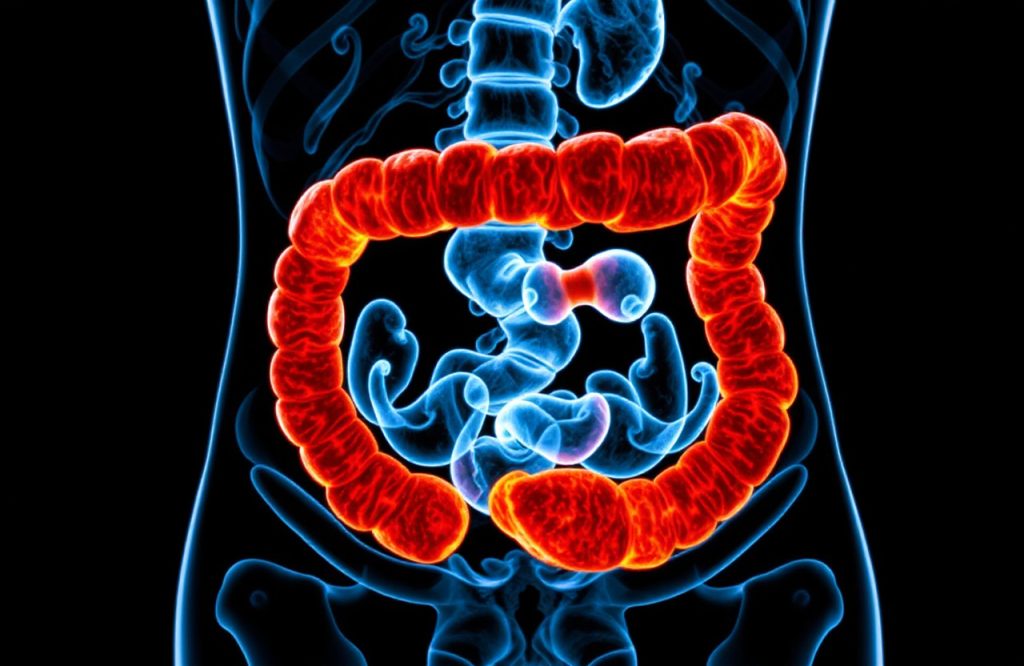
Understanding Inflammatory Bowel Disease (IBD)
IBD is characterized by chronic inflammation of the lower gastrointestinal tract and includes conditions such as Crohn’s disease and ulcerative colitis. Patients suffering from IBD must regularly monitor their condition due to ongoing flare-up symptoms and an elevated risk of developing colon cancer.
Comparison with Traditional Diagnostic Methods
Traditionally, IBD diagnosis relies on endoscopy, an invasive procedure that does not provide detailed information on specific immune cell markers. An optimal diagnostic method would be less invasive, ensuring patient comfort while also allowing for accurate diagnosis in cases where inflamed areas of the gastrointestinal tract are not visible through endoscopy.
Application of Immuno-PET in Ulcerative Colitis Diagnosis
In this research, Immuno-PET imaging was employed to evaluate mice with ulcerative colitis, contrasting their results with those of healthy mice. The findings revealed that mice with ulcerative colitis exhibited a higher concentration of inflammatory markers in their colons, with increases ranging from three to five times more than their healthy counterparts. Additionally, magnetic resonance imaging (MRI) results indicated a notable rise in inflammation signals in the affected mice compared to healthy ones. Notably, a correlation was observed between the heightened inflammation detected by Immuno-PET and percentage body weight loss, a relationship not evident with MRI.
Implications for the Future of IBD Diagnosis and Treatment
The study concluded that Immuno-PET outperforms MRI in identifying inflammation markers, thereby facilitating more efficient diagnosis and monitoring of IBD. Most treatments for IBD focus on specific inflammatory markers, and this new technology could potentially predict the effectiveness of these therapies without the need for invasive endoscopic procedures.
Conclusion
This breakthrough in diagnostic imaging signifies a substantial advancement in the management of inflammatory bowel disease, potentially improving patient outcomes through timely and less invasive evaluations.
References
Ng SC, et al. Worldwide incidence and prevalence of inflammatory bowel disease in the 21st century: a systematic review of population-based studies. Lancet. 2018 Dec 23;390(10114):2769-2778. doi: 10.1016/S0140-6736(17)32448-0.
Society of Nuclear Medicine and Molecular Imaging. Immuno-PET precisely diagnoses IBD inflammation without invasive procedures. EurekAlert. 17-JUN-2019.
Image by David Mark from Pixabay.