From Lab to Clinic: A Novel Test for Endometriosis
Introduction
This article is part of our Special Series “From Lab to Clinic.” For those interested in following the progress of this innovative test through clinical trials and regulatory approvals, we invite you to stay updated with us here.
Promising Preliminary Results
In March 2024, researchers from Oxford University revealed encouraging preliminary findings from the first non-invasive diagnostic test for endometriosis. The DETECT (Detecting Endometriosis expressed integrins using technetium-99m) imaging study is currently being conducted in Oxford, United Kingdom. The trial, led by Professor Christian Becker, Co-Director of the Oxford Endometriosis CaRe Centre, and Krina Zondervan, Head of the Nuffield Department of Women’s and Reproductive Health, aims to recruit between 20 and 25 women, with hopes of completing this phase of the study by the end of 2024.
Encouraging Early Results
Initial results indicate that a novel imaging system, akin to a CAT scan, can successfully detect early stages of peritoneal endometriosis. In one instance, researchers identified a lesion that had been missed during ultrasound testing, which was later confirmed through surgical intervention. Dr. Tatjana Gibbons presented these findings at the Society for Reproductive Investigation (SRI) annual meeting in March 2024, held in Vancouver, Canada.
Track and Trace Technology
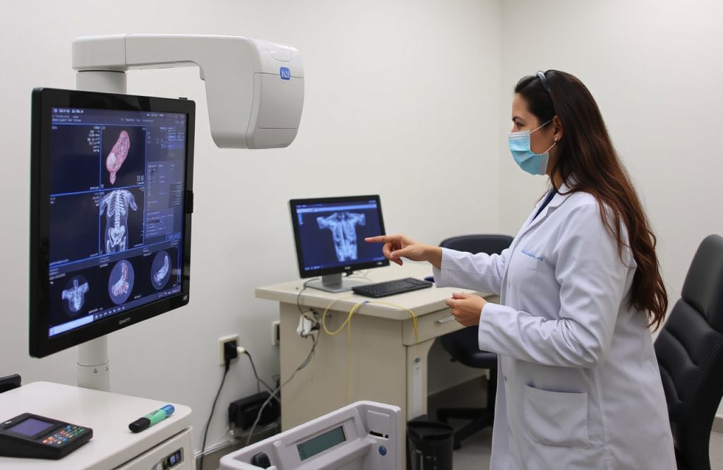
The imaging technology employs a radio-labeled tag to mark developing blood vessels. This tag effectively identified clusters of endometrium-like tissue that had developed outside the uterus. Similar to a thyroid scan, the patient receives an injection of a radio-labeled tracer. As the tracer circulates, it adheres to any tissue with newly formed blood vessels. Subsequently, the patient undergoes scanning in a SPECT-CT body scanner, which utilizes a sensitive camera to detect the faint radioactive signals emitted by the tracer.
First Visualization of Endometriosis
For the first time, researchers were able to visualize endometriosis within the peritoneum, the lining of the pelvis and abdomen. This discovery is significant, as visualizing this condition typically requires surgical intervention. The most prevalent form of endometriosis can currently only be diagnosed through laparoscopic (keyhole) surgery. Additionally, the study mapped endometrial lesions on internal organs, including the bladder, bowel, and ovaries.
SPECT-CT: A New Hope for Diagnosis
On average, it takes approximately seven years for a patient to receive a diagnosis of endometriosis. This lengthy delay is primarily due to the necessity for surgery under general anesthesia for diagnosis. If this new technology is adopted in hospitals, it could represent a significant advancement for women suffering from chronic abdominal and pelvic pain. As Professor Becker noted in a press release, “Endometriosis is a common disease affecting many millions of women worldwide with pain and infertility. The current delay in diagnosis results in prolonged suffering and uncertainty. Therefore, a novel imaging tool to assist healthcare professionals in identifying or ruling out the disease is urgently needed.”
Further Information
For more details, the SRI abstracts are available here.