A Comprehensive Meta-Analysis on MRI Imaging Data in Epilepsy Patients
Understanding Epilepsy
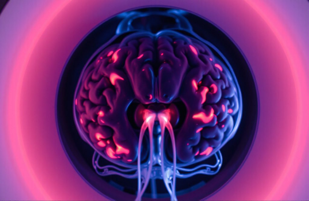
Epilepsy is a complex neurological disorder marked by seizures, which can range from subtle and barely noticeable to intense and involuntary shaking. This condition affects nearly 40 million individuals globally. Seizures result from abnormal electrical activity in nerve cells within the brain. While a small percentage of epilepsy cases are linked to genetic defects or significant brain trauma, such as injury or stroke, the majority remain idiopathic, with undetermined causes. Treatment often involves anti-convulsant medications, but responses vary; some patients may no longer need medication, while others may not respond at all, complicating diagnosis and management due to the disease’s variability.
Impact of Epilepsy on Brain Function
Epilepsy encompasses various types, each potentially affecting different regions of the brain’s cortex. Researchers employ techniques such as magnetic resonance imaging (MRI) and brain tissue analysis, the latter requiring post-mortem samples since biopsies are not routinely conducted on living patients. While MRI scans can reveal pathological markers of epilepsy, the extensive imaging data collected globally has yet to be systematically compiled and analyzed. Understanding the distinct or shared disease markers among different epilepsy forms could enhance therapeutic targeting and promote personalized treatment approaches.
The ENIGMA Study
Overview of the Study
The ENIGMA (Enhancing Neuro Imaging Genetics through Meta-Analysis) study, recently published in the journal BRAIN, stands as the largest neuroimaging analysis of epilepsy to date. This comprehensive research involved contributions from 24 research centers across 14 countries, including regions in Europe, North and South America, Asia, and Australia. Previous large-scale studies have highlighted structural brain abnormalities in various neurological disorders, including schizophrenia, depression, and obsessive-compulsive disorder.
Research Objectives
The researchers aimed to achieve several critical goals through this meta-analysis:
1. Investigate whether distinct types of epilepsy exhibit similar structural brain abnormalities.
2. Examine mesial temporal lobe epilepsy (MTLE) for differences between individuals affected on either side of the brain.
3. Analyze idiopathic generalized epilepsies (IGE), believed to have a genetic basis, which often show normal MRI results.
The study compiled imaging data from 2,149 epilepsy patients and 1,727 healthy controls, enabling rigorous statistical analysis.
Key Findings
In their first analysis, researchers discovered that various types of epilepsy displayed common structural anomalies across multiple brain regions, indicating that different disease types may share a neuroanatomical signature. In the second analysis, they observed that individuals with right-sided MTLE did not show damage to the left hippocampus and vice versa. Interestingly, damage extended beyond the hippocampus, suggesting that MTLE might operate as a network disease rather than a strictly localized one. Lastly, in the analysis of IGE, contrary to previous assertions of normal MRI findings, several structural irregularities were identified, including reduced brain volume and thickness in multiple areas.
Conclusion: Advancing Epilepsy Research
The authors acknowledged limitations in their study, such as the reliance on cross-sectional data, which does not clarify whether observed features contributed to past brain damage or resulted from progressive trauma. Additionally, the study could not fully account for factors like medications, seizure types, frequency, and disease severity. Nonetheless, this extensive meta-analysis marks a significant advancement in our understanding of how different forms of epilepsy impact the brain and holds promise for the development of more personalized and effective treatment strategies.
Written by Adriano Vissa, PhD
Reference: Whelan CD, et al. Structural brain abnormalities in the common epilepsies assessed in a worldwide ENIGMA study. Brain. 2018; 141(2):391-408