Understanding MRI and CT Scans
What is an MRI and How Does it Work?
MRI, or magnetic resonance imaging, is a medical imaging technique that produces detailed images of the body, including cross-sections of organs and tissues. This method utilizes a combination of radio waves and strong magnetic fields to generate highly accurate diagnoses for conditions such as diseases, strokes, and tumors. The process involves the reorientation of subatomic particles found in the body’s water molecules, allowing for precise imaging.
Overview of an MRI Appointment
Preparing for an MRI appointment is relatively simple. Patients must avoid metallic objects and accessories due to the powerful magnetic field. Individuals with metallic implants should consult their physician about the safety of undergoing an MRI. During the scan, it is crucial for patients to remain still, as movement can affect image quality. Although the machine produces loud noises, patients are often provided with headphones or earplugs for comfort.
Impact of MRI on the Progression of Science
MRI technology has made significant contributions to medical science, offering a non-invasive approach that is typically painless with minimal side effects. Notably, MRI screenings have demonstrated greater sensitivity in detecting breast cancer compared to mammograms. Additionally, MRI is advantageous in brain tumor segmentation and is recognized as the most accurate method for assessing heart ventricle function. It also plays a vital role in evaluating chronic liver disease while avoiding the risks associated with ionizing radiation, and is effective in categorizing male infertility.
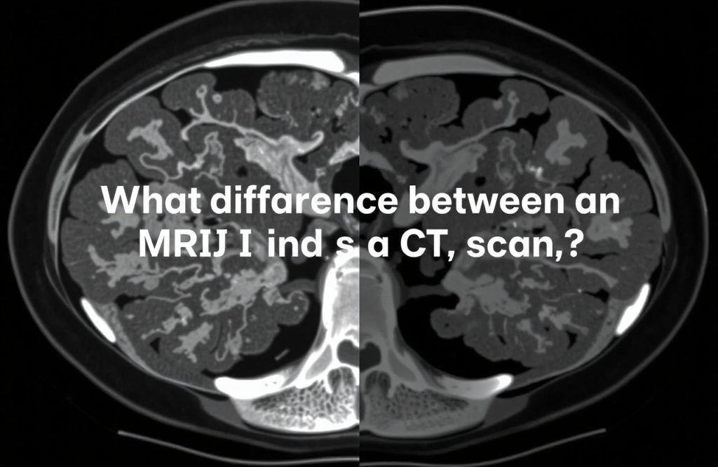
What is a CT Scan and How Does it Work?
A CT scan, or computerized tomography scan, employs x-rays to generate intricate cross-sectional images of various body parts, including blood vessels. By combining multiple images taken from different angles, CT scans excel in diagnosing internal injuries, tumors, and diseases.
Concerns about CT Scans and Cancer
One significant distinction between MRI and CT scans is that CT scans use high-frequency x-rays, which can expose patients to harmful ionizing radiation. Recent studies have raised concerns regarding the potential cancer risk associated with CT scans. Although a single CT scan presents a low risk, the danger increases with multiple scans. Efforts to reduce radiation exposure, particularly in pediatric radiology, remain complex and require ongoing innovation.
Overview of a CT Scan Appointment
Before a CT scan, patients must remove all metal objects and stay still during the imaging process. The scanner captures x-rays from various angles, which are processed by computer software to create cross-sectional images. Unlike MRI, CT scans can be more invasive; in certain cases, patients may need to fast and receive contrast material via injection or oral administration for clearer imaging.
Renowned Uses of CT Scans in Medical Sciences
CT scans provide medical professionals with valuable insights similar to MRI. They are particularly effective in assessing spine and hip fractures in individuals over 65 and have advanced the understanding of internal bleeding from torso trauma. Additionally, CT scans aid in diagnosing rare intracranial tumors and facilitate guided needle injections into joints through a combination of real-time ultrasonography and CT imaging. They are also instrumental in constructing three-dimensional images of human blood vessels.
How Do These Imaging Methods Compare in Contemporary Medicine?
While CT scans have traditionally been the standard for diagnosing acute strokes, research indicates that MRI is now more effective for emergency diagnoses in typical patient samples. Furthermore, MRI has proven superior in detecting chronic bleeding within brain tissue. However, CT scans still have advantages, particularly in diagnosing certain mediastinal tumors in the chest. Both MRI and CT technology offer remarkable imaging capabilities, each with unique strengths in medical diagnostics.